








-
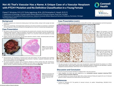
Not All That’s Vascular Has a Name: A Unique Case of a Vascular Neoplasm with PTCH1 Mutation and No Definitive Classification in a Young Female
Casey P. Schukow, Emily Jagenburg, and Christopher K. Hysell
Publication Date: 4-15-2026
Background ▪ Vascular neoplasms are a broad and complex group of soft tissue entities, arising in both younger and older populations. ▪ While tumor morphology, immunophenotypic patterning, and clinical correlation are essential in establishing accurate diagnoses, molecular profiling has increasingly become a vital tool in deciphering challenging cases. Case Presentation ▪ We present a case of a female in her mid-twenties who presents with a shoulder mass that was detected on imaging (outside institution, image not available). Core biopsy of the mass showed a vascular neoplasm most compatible with retiform hemangioendothelioma, which is an uncommon entity with propensity to locally recur unless wide local excision is performed. ▪ Surgical resection was performed, and the gross specimen showed a fleshy, well-demarcated soft tissue lesion (4.4 cm greatest dimension
-
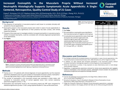
Increased Eosinophils in the Muscularis Propria Without Increased Neutrophils Histologically Supports Symptomatic Acute Appendicitis: A SingleCentered, Retrospective, Quality-Control Study of 21 Cases
Casey P. Schukow, Madison Konja, Zhenhong Qu, and Ping L. Zhang
Publication Date: 4-15-2026
Background ▪ The presence of neutrophils in muscularis propria is well known to correlate clinically with symptoms of acute appendicitis. ▪ Eosinophils present in the muscularis propria were noted in cases of acute appendicitis in 2000 (Carr, 2000), but the significance of their presence has been unclear, with limited supportive literature. ▪ The goal of this study was to investigate whether increased eosinophils in muscularis propria, without increased neutrophils, could be correlated with clinically diagnosed (symptomatic) acute appendicitis.
-
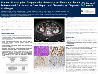
Chronic Consumptive Coagulopathy Secondary to Metastatic Poorly Differentiated Carcinoma: A Case Report and Discussion of Diagnostic Challenges
Casey P. Schukow, Kaylyn Lilly, Ping L. Zhang, Marc D. Smith, and James Z. Huang
Publication Date: 4-15-2026
Introduction • Chronic consumptive coagulopathy is a persistent imbalance between clotting and bleeding, often triggered by underlying conditions like malignancy. • It involves excessive activation of coagulation pathways and depletion of platelets and clotting factors. • One important differential is paraneoplastic hyperfibrinolysis, which is a rare condition where tumor cells excessively activate fibrinolytic systems. • Due to limited literature, this case aims to highlight diagnostic complexities and differentials.
-
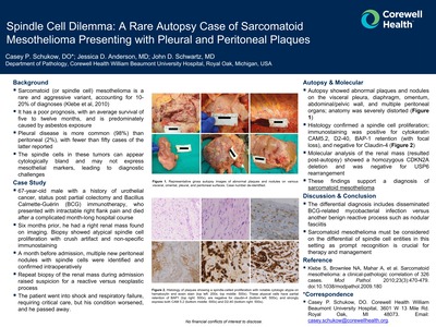
Spindle Cell Dilemma: A Rare Autopsy Case of Sarcomatoid Mesothelioma Presenting with Pleural and Peritoneal Plaques
Casey P. Schukow, Jessica D. Anderson, and John D. Schwartz
Publication Date: 4-15-2025
- Sarcomatoid (or spindle cell) mesothelioma is a rare and aggressive variant, accounting for 10- 20% of diagnoses (Klebe et al, 2010)
- It has a poor prognosis, with an average survival of five to twelve months, and is predominately caused by asbestos exposure
- Pleural disease is more common (98%) than peritoneal (2%), with fewer than fifty cases of the latter reported
- The spindle cells in these tumors can appear cytologically bland and may not express mesothelial markers, leading to diagnostic challenges
-
Lipoid Pneumonia Due to Ingested and Aspirated Olive Oil Presenting as a Right Middle Lobe Mass Lesion in an Elderly Male with Chronic Reflux Disease: An Unusual Mimicker of Malignancy
Casey P. Schukow, Said Khayyata, and Barbara S. Ducatman
Publication Date: 4-16-2025
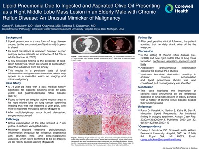
-Lipoid pneumonia is a rare form of lung disease caused by the accumulation of lipid (or oil) droplets in alveoli - Its exact prevalence is unknown; however, a prior autopsy series estimate an incidence of 1-2.5% in adults (Rana et al, 2020) - A key histologic finding is the presence of lipidladen histiocytes, which are unable to successfully clear the substance from the airway -This results in a persistent state of local inflammation and granuloma formation, which may appear as a mass-like lesion on imaging and mimic malignancy
-
Nocardia: An Unsuspecting Culprit Identified in the Brain Abscess of an Elderly Immunosuppressed Male
Casey P. Schukow and Yelena Wilson
Publication Date: 4-16-2025
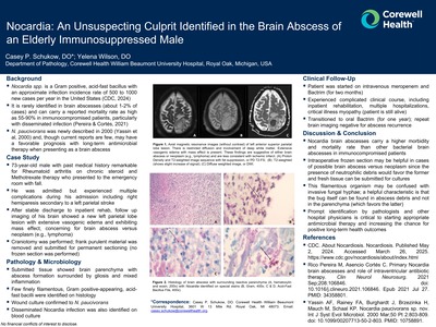
Background: Nocardia spp. is a Gram positive, acid-fast bacillus with an approximate infection incidence rate of 500 to 1000 new cases per year in the United States (CDC, 2024). It is rarely identified in brain abscesses (about 1-2% of cases) and can carry a reported mortality rate as high as 55-90% in immunocompromised patients, particularly with disseminated infection (Pereira & Cortés, 2021). N. paucivorans was newly described in 2000 (Yassin et al, 2000) and, though current reports are few, may have a favorable prognosis with long-term antimicrobial therapy when presenting as a brain abscess
-

Feeling Something Deeper: A Case of Hyalinizing Clear Cell Carcinoma of Lung Primary With Suspected Lynch Syndrome
Hussein Bazzy, Cameron Hubbard, Ahmad Tahawi, Narayana Gandham, Richard Zekman, Phillip V. Kaplan, Erich J. Schwartz, and Kevin Jamil
Publication Date: 5-22-2024
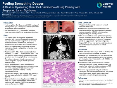
Hyalinizing Clear Cell Carcinoma (HCCC) is a type of Salivary Gland-type Tumor (SGT) that can present as a rare subset of lung neoplasms1
The association of HCCC with loss of mismatch repair expression (MMR) has not yet been described.
-

Using an Artificial Intelligence (AI) Tool (ChatGPT) to Identify the Main Indications and Side Effects of Drugs
Mai Elzieny, Christian Santiago, and Zhenhong Qu
Publication Date: 7-2024
This study evaluated the efficacy of ChatGPT, an AI powered language model (1), in retrieving drug information for pathology practice compared to UpToDate. The research focused on 160 commonly prescribed drugs, assessing the speed and accuracy of information retrieval. Results showed that ChatGPT efficiently provided indications and side effects of multiple drugs in batches within seconds, comparable to UpToDate's information but remarkably faster (2). The study suggests that ChatGPT is a promising valuable tool to help pathologists in accessing drug information, potentially improving diagnostic accuracy and work efficiency
-

CD21 Stains Dendritic Networks in Tertiary Lymphoid Structure of Transplant Renal Biopsies to Avoid Misdiagnosis of Borderline Changes
Brandon D. Metcalf, Wei Li, Hassan Kanaan, and Ping L. Zhang
Publication Date: 5-9-2024
Tertiary Lymphoid Structures (TLS) have been described as germinal center-like lymphoid structures composed dendritic network, mixed lymphocytes and high endothelial venules in internal organs following primary lymphoid organs (thymus and spleen) and secondary lymphoid structures (lymph nodes and native lymphoid structures along GI tract).
The TLS is mainly located at peri-glomeruli, peri-vascular areas or renal pelvis following inflammatory insults. A recent study indicates that TLS can been seen up to 19 % of protocol transplant renal biopsies (TRB) (JASN, 2022;33:186-200).
We hypothesize that CD21 staining can identify dendritic cells of TLS so that borderline changes are not over-diagnosed.
-

Optimize Artificial Intelligence Language Model Use in Medical Board Exams: Insights From Instruction Quality and Domain Context Analysis
Zhenhong Qu, Mai Elzieny, and Kanika Arora
Publication Date: 7-2024
Artificial intelligence language models (AI-LMs) show promise in medical education and clinical problem solving, but their performance in medical board exams has been inconsistent (1,2,3).
This study investigated how prompt engineering and providing essential reference material influence AI-LM performance on board exam questions.
We tested ChatGPT and Claude-2 on 360 questions in various formats under three conditions: simple prompt, elaborate CRAFTS prompt, and CRAFTS prompt with references (CRAFTS stands for context, role, action, format, tone, and style).
Results showed significant improvement in accuracy from simple prompts (67-83.5%) to CRAFTS prompts (76.2-92.5%) to CRAFTS prompts with references (97.3- 99.4%)
These findings suggested that AI-LM performance in medical exams is heavily influenced by instruction quality and provided context, highlighting the need for standardized evaluation methods.
-

The Severity of Obstructing Tubulopathy is Often Associated with Foci of Renal Tubular Obstruction
Ping L. Zhang, Hassan D. Kanaan, and Wei Li
Publication Date: 10-2024
Etiologies of renal failure due to renal tubular injury may result from either ischemic/toxic injury of proximal tubules (ATN), variants of interstitial nephritis or obstructing tubulopathy by the luminal blocking of either monoclonal casts, uric acid or calcified deposits. Our previous studies indicate that the polarizable semi-transparent calcium oxalate deposits block proximal tubules, whereas monoclonal casts, uric acid deposits and bluish calcium phosphates block the distal tubules. We hypothesized that the serum creatinine (SCr) levels and the number of tubular obstructing deposits were correlated so that pathologic identification of obstructing foci can be predictable for the extent of renal dysfunction.
-

Grading and Follow-Up of T Lymphocyte-Mediated Acute Interstitial Nephritis Following Checkpoint Inhibitor Therapy
Ping L. Zhang, Wei Li, and Hassan D. Kanaan
Publication Date: 10-2024
Immuno-check point inhibitors (CPI) such as PD-1/PD-L1 inhibitors have been used to treat a variety of metastatic carcinoma with positive effects. However, there are complications such as gastrointestinal, hepatic and endocrine symptoms and kidney dysfunction. We describe our experience with CPI-associated acute interstitial nephritis (AIN).
-

Effect of Comorbidities and Choice of Treatment on Overall Survival in Elderly Patients with Acute Myeloid Leukemia: A Beaumont Experience
Bilal M. Ali, Emma Herrman, James Huang Huang, and Mohammad Muhsin Chisti
Publication Date: 5-2023
First line therapy for Acute Myeloid Leukemia (AML) is 7+3 regimen. It often cannot be used in elderly patients due to intensity. Venetoclax + hypomethylating agent (HMA) is approved for AML treatment in these patients. We investigate the efficacy of this treatment in a community setting where patients do not have the same resources available to them as a large academic center. Primary outcome was survival of patients greater than 60 years of age with a diagnosis of AML who received 7+3 therapy versus those who received venetoclax + HMA. Secondary outcomes included characteristics of those who received the two therapies.
-
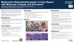
Multicentric Reticulohistiocytosis: A Case Report With Molecular Analysis and Discussion
Casey Schukow, Katherine Burke, Arivarasan Karunmurthy, and Jonhan Ho
Publication Date: 10-7-2023
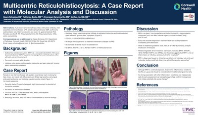
Multicentric reticulohistiocytosis (MRH) is a rare non-Langerhans cell histiocytosis that affects multiple organ systems, including the skin and joints. It commonly occurs in adult females and is diagnosed based on clinical presentation with associated radiologic and histologic findings. Histologic features include an infiltrate composed of multinucleated histiocytes and giant cells with a characteristic “ground glass” eosinophilic cytoplasm. Although the exact prevalence is unknown, current literature estimates that anywhere from 300 to 400 cases have been documented since being first reported in 1937. Here we present a case of MRH in a 45-year-old female who presented with eruptive red papules on the face, neck, back, hands and arms in addition to arthritis of the wrists and hands. A punch biopsy revealed dermal multinucleated histiocytes with ground glass cytoplasm positive for CD163, and negative for SOX10, S100, BRAF, and ALK. Additional immunostains and molecular testing via next-generation sequencing are pending at the time of abstract submission. BRAF, MAP2K1, TET2, KIF5B, FGRF1, and KRAS have been suggested in prior studies as potential genetic drivers, implicating MRH as a neoplastic versus autoinflammatory disease. However, overall literature is lacking. Thus, this report will provide an updated discussion regarding the molecular underpinnings of MRH. Furthermore, the authors will examine distinguishing genetic and histopathologic differences of relevant MRH mimickers to inform practicing pathologists.
-
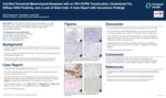
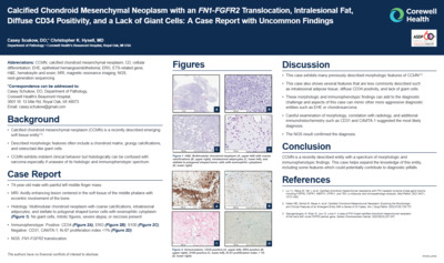
Calcified Chondroid Mesenchymal Neoplasm With an FN1-FGFR2 Translocation, Intralesional Fat, Diffuse CD34 Positivity, and a Lack of Giant Cells: A Case Report With Uncommon Findings
Casey Schukow and Christopher K. Hysell
Publication Date: 10-7-2023
Calcified chondroid mesenchymal neoplasm (CCMN) is a term for an emerging group of soft tissue tumors that display cartilaginous matrix formation and harbor translocations involving the FN1 gene. These tumors are rare and behave indolently but can be mistaken for malignant neoplasms. Current literature indicates that these tumors frequently contain giant cells, and infrequently contain intralesional fat or stain for CD34. Henceforth, we present a case of CCMN with uncommon findings causing a soft tissue finger mass in a 74-year-old male with some erosion into the underlying bone. Microscopically, the mass was composed of a multinodular chondroid neoplasm with areas of abundant coarse calcifications. Tumor cells were polygonal to stellate with eosinophilic cytoplasm, and interspersed adipocytes were present within the lesion. Giant cells were absent. Immunohistochemically, the tumor was diffusely positive for CD34, as well as ERG and S100. Next-generation sequencing confirmed the presence of an FN1-FGFR2 translocation and the diagnosis of CCMN. While this case showed many features that have been previously described in the literature, this case also showed some aspects that are uncommon based on the literature and could add to diagnostic difficulty. These include CD34 staining, which in conjunction with ERG staining could raise the possibility of epithelioid hemangioendothelioma, and interspersed adipocytes, which could heighten concern for an infiltrating malignant neoplasm like chondrosarcoma. The findings in this presented case help continue to expand the knowledge of an emerging subcutaneous pathologic entity, and highlight some uncommon features that should not dissuade one from making a diagnosis of CCMN.
-
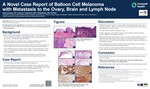
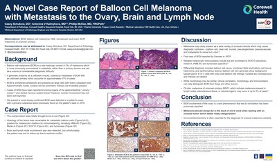
A Novel Case Report of Balloon Cell Melanoma With Metastasis to the Ovary, Brain and Lymph Node
Casey Schukow, Antonina V. Kalmykova, and Phillip McKee
Publication Date: 10-7-2023
Balloon cell melanoma (BCM) is a rare histologic variant (< 1%) of malignant melanoma characterized by large, foamy melanocytes with cytoplasmic vacuoles and pronounced nucleoli (“balloon cells”). Clinical presentation often varies and primary cutaneous lesions may be unknown in up to 47% of cases. Metastatic BCM tumors tend to display prominent balloon cell morphology, and cases have been reported involving organs of the gastrointestinal, urinary, ocular, and central nervous system tracts. However, ovarian involvement has not been well-reported. We present a novel biopsy-confirmed BCM case detected in a patient’s ovary with a primary cutaneous lesion previously found on the patient’s back in 2018. The ovarian lesion was initially thought to be a cyst, however the histology of the lesion was remarkable for metastatic balloon cells positive for melanocytic markers on immunostaining, including Melan-A, HMB45, SOX10, and tyrosinase. Brain and lymph node involvement was also detected, but unfortunately the patient was lost to follow-up due to wartime conflict and the remainder of the patient’s history is unknown. After conducting a thorough review of literature published on PubMed, the authors did not recognize previously reported cases of malignant BCM to the ovary to date. In this report, we recognize the importance of proper clinicohistologic and immunostain correlation when differentiating clear cell or xanthomatous lesions while briefly discussing how to differentiate these entities from BCM.
-
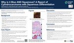
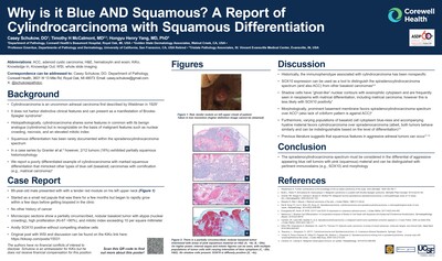
Why Is It Blue and Squamous? A Report of Cylindrocarcinoma With Squamous Differentiation
Casey Schukow, Timothy H. McCalmont, and Hongyu Henry Yang
Publication Date: 10-7-2023
Cylindrocarcinoma is an uncommon adnexal carcinoma first described by Wieldman in 1929. It does not harbor distinctive clinical features and can present as a manifestation of Brooke-Spiegler syndrome. Histopathologically, cylindrocarcinoma shares some features in common with its benign analogue (cylindroma) but is recognizable on the basis of malignant features such as nuclear crowding, necrosis, and an elevated mitotic index. Historically, the immunophenotype associated with cylindrocarcinoma has been nonspecific, but SOX10 expression can now be used as a tool to distinguish the spiradenocylindrocarcoma spectrum (and also transformed adenoid cystic carcinoma) from other basaloid carcinomas. Squamous differentiation has been rarely documented within the spiradenocylindrocarcinoma spectrum. In the series of Granter et al. (2000), however, 2/12 tumors (16%) exhibited partially squamous histomorphology. We report a poorly differentiated example of cylindrocarcinoma with marked squamous differentiation that mimicked other types of basaloid carcinoma with cornification, in particular matrical carcinoma. A specific diagnosis was possible on the basis of conventional microscopic features and SOX10 immunopositivity. The literature and differential diagnosis of keratinizing basaloid carcinomas will be reviewed.
-

Eosinophils Are a Useful Morphologic Marker to Scrutinize Plasma Cells in Endometrial Polyps
Olabisi Afolayan-Oloye, Jessica Anderson, and Ping L. Zhang
Publication Date: 9-2022
Chronic endometritis is characterized by presence of plasma cells, which can be confirmed by immunostaining for CD138. Endometrial polyps can irritate endometrium and harbor some plasma cells. As eosinophils are much easier to be identified than plasma cells on hematoxylin and eosin stained sections, the goal of this study was to investigate if the identification of eosinophils in endometrial polyps was a useful marker for identifying plasma cells.
-

Myeloperoxidase (MPO) Immunostaining Can Identify Endothelial Injury of Peritubular Capillaries and Glomeruli in Renal Antibody Mediated Rejection
Olabisi Afolayan-Oloye, Megan Moore, Wei Li, Hassan D. Kanaan, and Ping I. Zhang
Publication Date: 3-2022
-

CD30 Expression in Cutaneous B-Cell Lymphomas
Olabisi Afolayan-Oloye, Lili Zhao, Trilokraj Tejasvi, May P. Chan, Paul W. Harms, Douglas R. Fullen, Ryan A. Wilcox, and Alexandra C. Hristov
Publication Date: 10-2022
CD30 expression has been infrequently described in cutaneous B-cell lymphomas (CBCL). We examined CD30 expression in reactive lymphoid hyperplasia (RLH) and CBCL and correlated expression with clinicopathologic features.
-

Acute Pericarditis and Acute Pleuritis/Empyema Following Submandibular Infection in a COVID-19 Positive Patient - an Autopsy Revealing the Danger Space of the Neck
Jessica D. Anderson, Seyedalireza Fatemi, Joseph Fullmer, and Ping I. Zhang
Publication Date: 10-2022
Acute pericarditis and empyema are life-threatening complications of severe odontogenic infections; reports of these findings from an autopsy perspective are rare. We report an autopsy case demonstrating infection from the mandibular molars to the pericardium and pleura in a patient following Covid-19 infection
-

Varieties of Renal Diseases Identified in Renal Biopsies of Patients Infected by COVID-19
Jessica D. Anderson, Wei Li, Hassan D. Kanaan, and Ping L. Zhang
Publication Date: 10-10-2022
COVID-19 has been shown to cause renal pathology by multiple proposed mechanisms. However, studies evaluating renal biopsies for the effects of COVID-19 remain limited. We report our experience in variety of renal pathologic diagnoses caused by COVID-19 infection in our health system.
-

Spectrum of C3 glomerulopathies (C3GNs) – single center experience
Mustafa M. Deebajah, Wei Li, Hassan Kanaan, and Ping L. Zhang
Publication Date: 10-10-2022
C3GNs represent a rare type of glomerulopathy due to the activation of alternative complement pathway, including C3 dominant glomerulonephritis (C3D-GN) and dense deposit disease (DDD). The goal of this study was to demonstrate a spectrum of C3GNs.
-

IgG4-Related Interstitial Nephritis (IRIN) Ranges from Kidney Mass to Acute Kidney Injury (AKI)
Mai Elzieny, Wei Li, Hassan D. Kanaan, and Ping L. Zhang
Publication Date: 10-2022
IRIN can present as either renal mass or with AKI. The goal of the study was to present our 4 IRIN cases with different clinical scenarios.
-

Anti-NMDA-Receptor Encephalitis in a Patient with Ovarian Teratoma, Harboring Brain Histology of Varying Developmental Stages
Seyedalireza Fatemi, Elaine Qu, and Joseph Fullmer
Publication Date: 6-2022
Anti-NMDA-receptor encephalitis is a subacute, autoimmune disorder thought to be caused by autoantibodies directed against the N-methyl-D-aspartate (NMDA) receptor. Although NMDAR encephalitis is a familiar entity to psychiatrists and neurologists, it is less commonly reported in the pathology literature. Clinical symptoms of anti-NMDAR encephalitis may mimic schizophrenia and psychotic spectrum disorders or substance-induced psychosis. Although initially described in association with ovarian teratomas in women, anti-NMDAR encephalitis has been reported in individuals without paraneoplastic association, as well as in males. Previous literature has suggested NMDA receptor expressing teratoma neurons are densely aggregated and are smaller in size. Ki-67 index can be higher in these neurons and they usually show B-cell lymphocytic infiltration around them. Herein, we report a case of a 29-year-old woman with suicidal ideation and other neuropsychiatric manifestations who was found to have a right ovarian cystic mass by imaging study. Microscopically, the resected ovarian mass is composed of mature skin, fat, cartilage and neural tissues. Nerve, ganglions and multiple brain tissues are present. Interestingly, cerebellum including external granular cell layer (normally only seen in infants), cerebrum-like, choroid plexus and other neural elements are present. There is peripheral lymphoplasmacytic infiltrates around and within the neuroglial matrix. Cerebral spinal fluid tested positive for Anti-NMDAR. The combined clinical, histological, and laboratory findings confirmed the above diagnosis. Its resultant relationship to cystic teratoma warrants awareness of this condition by pathologists. Although slow to respond to treatment, this patient now continues to show improvement with plasmaphoresis. Portions of this abstract have been previously presented at the ASCP meeting in 2021.
Printing is not supported at the primary Gallery Thumbnail page. Please first navigate to a specific Image before printing.