


Files
Download Full Text (659 KB)
Description
Historically, myotonic dystrophy, type 1 (DM1), was thought to occur in 5-20 per 100,000 births, but recent studies suggest that the incidence may be as high as 1 per 2,100 births. Congenital DM1 (cDM1) has a variable reported incidence and is often the first familial diagnosis. cDM1 is often diagnosed in the neonatal period. There are certain ultrasound findings that support this diagnosis, such as fetal talipes and polyhydramnios, but none are pathognomonic. This case emphasizes how maternal and fetal diagnoses can coincide and that efficient diagnoses inform care for both patients.
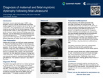
This case reviews a 29-year-old G1P0 who initially presented to the Maternal Fetal Medicine (MFM) for fetal ultrasounds due to suspected bilateral talipes, first noted at 20 weeks gestation. At her 25-week ultrasound, no fetal flexion or extension of legs was noted. Subsequently, a 29-week ultrasound demonstrated the bilateral lower extremities fixed in an extended "pike" position with minimal fetal movement. No other significant ultrasound findings were noted at that time. The patient also reported her own muscular symptoms, including muscle stiffness and gait difference, and the "Gowers's maneuver" was noted in the office. The patient was urgently referred to a medical geneticist. Her myotonia gene panel demonstrated >200 repeats in one copy of her DMPK gene, consistent with DM1. Following maternal diagnosis, the patient underwent amniocentesis for fetal genetic testing, which revealed an expanded DMPK allele with approximately 1710 repeats, consistent with cDM1.
During pregnancy, the patient connected with multiple subspecialists. She continued following with MFM and planned for a delivery at a regional birthing center for maternal intrapartum cardiac monitoring and neonatal acute care services. She was ultimately scheduled for an amnioreduction followed by an induction of labor secondary to polyhydramnios at 37 weeks 2 days gestation. Fetal status was reassuring during labor, and her delivery was uncomplicated. The neonatal ICU was present at delivery. The umbilical cord was immediately clamped and cut due to nonreassuring neonatal status. APGARs were 3 and 5 at 1 and 5 minutes, respectively, due to poor tone and minimal reactivity. The baby was intubated during initial resuscitation and admitted to the neonatal ICU for 60 days, requiring extensive multidisciplinary care. They were ultimately discharged with oxygen support but without a tracheostomy. Both the patient and baby continue to follow with subspecialists routinely.
This case supports second- and third-trimester ultrasound findings associated with cDM1, while exemplifying how fetal findings can expedite the often delayed maternal diagnosis. Further, it demonstrates the importance of expeditious diagnosis to establish surveillance and management of maternal and neonatal complications.
Publication Date
5-8-2026
Disciplines
Obstetrics and Gynecology
Recommended Citation
Bright V, Ismailova I, Fricke E. Diagnosis of maternal and fetal myotonic dystrophy following fetal ultrasound. Presented at: Research Day Corewell Health West; 2026 May 8; Grand Rapids, MI.
Comments
2026 Research Day Corewell Health West, Grand Rapids, MI, May 8, 2026. Abstract 2032