


-

Predicting Coronary Artery Calcium Using Abdominal Aorta and Visceral Artery Calcification
Jared Dixon, Sayf Al-Katib, Kiran Nandalur, Jacob Ghannam, Ali Beydoun, and Desiree Clement
Publication Date: 5-2025
CAC scoring has emerged as a widely available, consistent, and reproducible means of assessing risk for major cardiovascular outcomes, especially useful in asymptomatic people for planning primary prevention interventions such as statins and aspirin.1 The Multiethnic Study of Atherosclerosis (MESA) demonstrated that a CAC score of 0 represents a less than 5 percent 10-year Atherosclerotic cardiovascular disease (ASCVD) risk. CAC scores 1-299 represent borderline to intermediate risk, and CAC score >300 represents a “high risk” 10 percent or greater 10-year ASCVD risk.2
When used correctly, CAC scoring is a powerful predictor of ASCVD risk and holds the key for many patients to receive appropriate and timely treatment for cardiovascular disease. Unfortunately, because testing is recommended only in patients that are asymptomatic for CVD, many of those to whom the screening test would be beneficial never have the test performed.3
This study aims to increase the reach of CAC scoring to a larger population prior to the onset of CVD symptoms by predicting CAC scores using a novel scoring system to grade calcification of the Abdominal Aorta (AA), Celiac (CA), Superior Mesenteric (SMA), and Renal Arteries (RA) discovered incidentally on abdominal non-contrast Computed Tomography imaging. Patients who receive abdominal imaging for kidney stone protocol, abdominal pain, or other reasons may also be screened for ASCVD risk.
In prior studies AA calcification has been well described as predictive of CAC scores4, 5, 6, but to our knowledge no studies have attempted to correlate the AVAC scores with CAC scores. A combination of both scores could be more predictive of CAC than AA scoring alone.
-

Jamestown Canyon virus meningoencephalitis: A case report
Amy Ishbia, Hannah Guider, Matthew Sierra, and Jonathan Doty
Publication Date: 3-13-2025
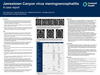
Jamestown Canyon virus is a rare mosquito-borne orthobunyavirus that typically is asymptomatic or causes mild symptoms. However, it can rarely cause severe neuroinvasive disease [2,5]. The primary hosts include white-tailed deer, although moose, elk, and bison can also be hosts [2].
Typically, cases are seen between April and September, with a bimodal distribution peaking in spring and late summer [2].
Number of cases in the U.S. in 2024: 16 total cases, 11 neuroinvasive [1]. Number of cases in the U.S. between 2011-2023: 308, with 10 deaths, 206 neuroinvasive. One case was reported from Alpena, MI, where our patient resided [1].
-

Prostatic Urethral Length on MRI as a Predictor of Late Genitourinary Toxicity After Prostate Cancer Radiation
Kyu Kim, Joseph Lee, Allison Hazy, Sayf Al-Katib, Hong Ye, Nathan Kolderman, Abhay Dhaliwal, Daniel Krauss, Thomas Quinn, Kimberly Marvin, and Kiran R. Nandalur
Publication Date: 5-2025
Radiation therapy (RT) is a widely used treatment for prostate cancer.1 While effective, some patients develop late genitourinary (GU) toxicity, which can impact quality of life.2,4 Identifying pre-treatment factors that predict toxicity risk may help improve clinical decision-making. Recent studies suggest that prostatic urethral length (PUL) seen on MRI is associated with an increased risk of late GU toxicity.3 Understanding this relationship could enhance risk stratification before treatment.
-

Advances in Minimally Invasive Hepatobiliary Interventions for Non-Operative Candidates
Tulasi Talluri, Philip Cieplinski, and Kristian Loveridge
Publication Date: 5-2025
Cholangioscopy is a technique used for diagnosing and treating patients with pathologies of the gallbladder, liver, and pancreas. It has been available since 1976 but with limited use in interventional radiology (IR) due to issues with steerability, irrigation capabilities, requirement for more than one operator, and poor percutaneous access options; however, direct intraluminal visualization of the gallbladder, common bile ducts, and even the ampulla and duodenum has become more practical with the advent of single operator percutaneous systems. Access to the biliary system can now be made through smaller 11 French sheath/delivery systems allowing for improved maneuverability into the common bile duct. The purpose of this study was to evaluate the efficacy and describe various applications of percutaneous cholangioscopy in patients with gallbladder and biliary stone duct disease who are ineligible for surgery and/or endoscopic retrograde cholangiopancreatography (ERCP).
-

Quantifying the Importance of Orthopaedic Surgeon Attributes by the Public
Rasheed Abdullah, James E. Feng, Phillip Vartanyan, Hassan Alosh, Drew Moore, Leonardo M. Cavinatto, and Betina Hinckel
Publication Date: 5-2024
Evaluate what surgeon attributes are currently important to our patients, and what modalities may be most effective in finding orthopaedic surgeons, using online crowdsourcing.
-

More Than You Can Swallow: Mycotic Aneurysm, An Uncommon Etiology of Dyspepsia
Hussein Bazzy, Katharine Glover, Steven Jones, James Aldridge, Michael Potes, and Katie Sumnicht
Publication Date: 5-9-2024
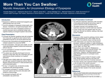
Dyspepsia is defined as predominant epigastric pain lasting greater than one month.
Common etiologies of dyspepsia include peptic ulcer disease, medications, and gastroesophageal reflux.
We present a rare case of dyspepsia caused by a mycotic aneurysm due to accidental foreign body ingestion leading to emergent inpatient intervention.
-

Feeling Something Deeper: A Case of Hyalinizing Clear Cell Carcinoma of Lung Primary With Suspected Lynch Syndrome
Hussein Bazzy, Cameron Hubbard, Ahmad Tahawi, Narayana Gandham, Richard Zekman, Phillip V. Kaplan, Erich J. Schwartz, and Kevin Jamil
Publication Date: 5-22-2024
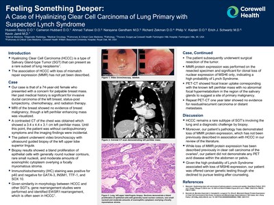
Hyalinizing Clear Cell Carcinoma (HCCC) is a type of Salivary Gland-type Tumor (SGT) that can present as a rare subset of lung neoplasms1
The association of HCCC with loss of mismatch repair expression (MMR) has not yet been described.
-

Varicella-Zoster Encephalitis Presenting with Trigeminal Neuralgia, Complicated by Vasculopathy
Amy Ishbia, Kathy Ross, Dillon Yaldo, Jacob Conroy, and Andrea Stoner
Publication Date: 5-9-2024
VZV is a herpesvirus that causes chickenpox before laying dormant and having the potential to reactivate to cause myelitis, encephalitis, or vasculopathy.
Encephalitis occurs in less than 0.1% of cases; however accounts for 90% of neurologic complications from VZV.
Vasculopathy is a rare complication of VZV encephalitis characterized by vessel wall damage and transmural inflammation with multinucleated giant cells and/or epithelioid macrophages.
Vasculopathy has significant morbidity/mortality due to ischemic/hemorrhagic stroke, spinal cord infarction, temporal artery inflammation, ischemic cranial neuropathies and cerebral venous thrombosis.
-

Nephrolithiasis Induced Hydronephrosis as an Unusual Pathology of Biliary Obstruction: A Case Report
Michaela Knaggs, Lianne Strimpel, Christian Przeslawski, Erika Michelin, and Ahmed Tahawi
Publication Date: 5-9-2024
We present an interesting case of biliary obstruction secondary to mass effect of hydronephrosis from nephrolithiasis managed jointly between general and urologic surgery.
-

You’re Testing My Patients: Clinicians’ Preferences for Structured Reporting and Measurement Data
Davit Melik, Adela Pouzar, Allan Brazier, and Sayf Al-Katib
Publication Date: 5-2024
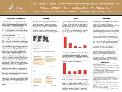
Radiological reports are a crucial part of patient care and management. Radiologists play a major role in patient care by interpreting imaging studies and making recommendations to referring physicians. 1,2,3 Although some physicians may prefer to initially interpret the imaging studies by themselves, radiologists’ reports have been shown to be significantly more accurate and detailed, which ultimately results in better patient care. 1,4,5,3 Thus it is important for radiology reports to be timely and as accurate as possible to improve overall patient care. The clarity of communication between radiologists and referring physicians in these reports remains an important factor in delivering quality care to patients.
Traditionally, radiology reports were created using free-text, narrative language. 1 In the last decade, however, radiology reports have transitioned from more traditionally formatted reports to a more highly structured format. 5 Structured reports in radiology are not a novelty and have been utilized since 2007 when they were first recommended by representatives of American College of Radiology. 3 The newer formats typically rely on specific templates, and they may contain disease-specific, system based or anatomical headers, as well as a structured description of normal and common abnormal findings. 4 Since structured reports started being widely used, different types of clinicians and other report “consumers” expressed specific preferences regarding the measurements and report styles they find more appropriate. Additionally, there are certain standardized reporting criteria (such as RECIST, WHO or SWOG) that are utilized in the field of radiology to report progression of specific types of pathologies (tumors in this case). 6,7 These criteria are generally well received by the scientific community and numerous studies support their implementation and use in day-to-day reporting. 7 However, some drawbacks are typically identified with them in evaluating the identification of disease progression. 7 To add to that, in some cases these criteria are not easily applicable in certain types of pathologies.
Some are concerned that structured reports will result in commodification of radiology and will hinder the freedom and the “art” of the practice. 11 While allowing radiologists to add free text where possible can help them maintain autonomy in reporting, national standardization guidelines can contain multiple specific templates created by broad and interdisciplinary consensus, with room for variation.
Ultimately, it is important to consider the expectations of all the clinicians involved in patient care, in order to maximize the level of care that patients receive. Particularly in patients with serious conditions such as cancer. The aim of this study will be to assess both report structure and quantitative data representation preferences of key lung cancer care clinicians at our institution.
-

Patient Population Representation in Medical Education Textbooks: Gender Imbalance of Diagnostic Images
Yasin Sahin, Abdul-Majid Khan, Nicolas Baker, Joshua Daniel, and Jacob Keeley
Publication Date: 5-2024
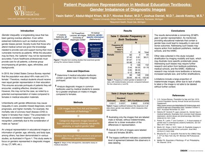
Gender inequality: a longstanding issue that has been gaining overdue attention. Even within esteemed institutions such as medical schools, gender biases persist. Society trusts that those who attend medical school are given the knowledge needed to provide care and support during their most vulnerable moments as patients. While the education may be there, the “patients” may not be represented accurately. Future healthcare professionals must provide care for all patients, a diverse group encompassing all genders, ages, ethnicities, and backgrounds.
In 2019, the United States Census Bureau reported that the population was about 49% male and 51% female.¹ Therefore, medical students should receive near equal gender representation in their education. This ensures accurate portrayals of patients they will encounter, enabling effective, directed care. However, this may not be the case, as noted by a potential overrepresentation of males compared to females in medical education materials.
Unfamiliarity with gender differences may cause inequality in care, possible missed diagnoses, worse prognoses, and higher mortality. For example, the mortality rate of coronary heart disease (CHD) is higher in females than males.2 The presentation for females is considered “atypical,” causing less recognition, potentially due to the consideration of males as “typical.”
An unequal representation in educational images or information of gender, age, ethnicity, and body type, among others, has been demonstrated in various studies, as illustrated in Figure 1. This study seeks to focus on genders represented in diagnostic images (X-ray, CT, MRI, etc.)
-

Invasive Lobular Carcinoma of the Breast Presenting as Pneumoperitoneum with Omental and Retroperitoneal Metastasis: A Case Report
Logan Smith, Christian Przeslawsk, Katie Sarraf, Steven Jones, and Raimundo Pastor
Publication Date: 5-9-2024
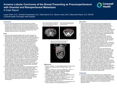
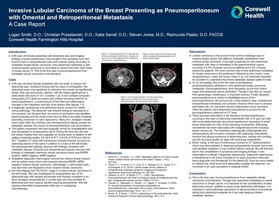
A 65 year old female presenting with abdominal pain and imaging findings of pneumoperitoneum was brought to the operating room and found to have a retroperitoneal mass and omental caking secondary to metastatic breast lobular carcinoma. Intra abdominal metastasis is rare however lobular carcinoma is more likely to spread here than other types of breast cancer. This is the first case of pneumoperitoneum from metastatic lobular carcinoma in the literature.
-

More Than You Can Swallow: Mycotic Aneurysm, An Uncommon Etiology of Dyspepsia
Hussein Bazzy, Katharine Glover, Steven Jones, James Aldridge, Michael Potes, and Katie Sumnicht
Publication Date: 5-4-2023
Dyspepsia is defined as predominant epigastric pain lasting greater than one month.
▪ Common etiologies of dyspepsia include peptic ulcer disease, medications, and gastroesophageal reflux.
▪ We present a rare case of dyspepsia caused by a mycotic aneurysm due to accidental foreign body ingestion leading to emergent inpatient intervention.
-

Varicella-Zoster Encephalitis Presenting with Trigeminal Neuralgia, Complicated by Vasculopathy
Amy Ishbia, Katherine Ross, Dillon Yaldo, Jacob Conroy, and Andrea Stoner
Publication Date: 5-4-2023
VZV is a herpesvirus that causes chickenpox before laying dormant and having the potential to reactivate to cause myelitis, encephalitis, or vasculopathy [1,4]. ▪ Encephalitis occurs in less than 0.1% of cases; however accounts for 90% of neurologic complications from VZV [2].
Vasculopathy is a rare complication of VZV encephalitis characterized by vessel wall damage and transmural inflammation with multinucleated giant cells and/or epithelioid macrophages.
Vasculopathy has significant morbidity/mortality due to ischemic/hemorrhagic stroke, spinal cord infarction, temporal artery inflammation, ischemic cranial neuropathies and cerebral venous thrombosis [2].
-

Impact of Structured Reporting Template on the Quality of HRCT Radiology Reports for Interstitial Lung Disease
Han G. Ngo, Girish B. Nair, and Sayf Al-Katib
Publication Date: 5-2023
This QI study compared the completeness of HRCT radiology reports before and after the implementation of a disease-specific structured reporting template for suspected cases of interstitial lung disease (ILD).
-

Complex Removal of Heterogenous Soft Tissue Mass in Patient with Prior History of Malignancy
Yumna Siddiqui and Randy Semma
Publication Date: 5-4-2023
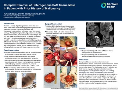
There is a variety of pathologies seen in the foot and ankle. One of the more challenging pathologies involves the ability to obtain the correct diagnosis and therapeutic treatment for a soft tissue mass of unknown etiology. With both benign and malignant tumors found in the lower extremities, most malignancy presenting in the foot and ankle is primary in nature. However, in patients with history of malignancy elsewhere in the body, a possible secondary metastatic tumor should always be a differential. Therefore, early diagnosis and treatment is of upmost importance. We present a case study of a patient with prior history of uterine cancer, presenting with an enlarging heterogenous soft tissue mass on her ankle.
-

Invasive Lobular Carcinoma of the Breast Presenting as Pneumoperitoneum with Omental and Retroperitoneal Metastasis A Case Report
Logan Smith, Christian Przeslawski, Katie Sarraf, Steven Jones, and Raimundo Pastor
Publication Date: 5-5-2023
A 65 year old female presenting with abdominal pain and imaging findings of pneumoperitoneum was brought to the operating room and found to have a retroperitoneal mass and omental caking secondary to metastatic breast lobular carcinoma. Intra abdominal metastasis is rare however lobular carcinoma is more likely to spread here than other types of breast cancer [1]. This is the first case of pneumoperitoneum from metastatic lobular carcinoma in the literature.
-

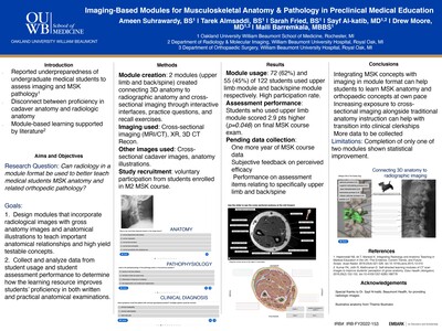
Imaging-Based Modules for Musculoskeletal Anatomy & Pathology in Preclinical Medical Education
Ameen Suhrawardy, Tarek Almsaddi, Sarah Fried, Sayf Al-katib, Drew Moore, and Malli Barremkala
Publication Date: 5-2023
Many medical students report a lack of adequate orthopedic and musculoskeletal (MSK) teaching in the preclinical medical curriculum. As gross anatomy is emphasized in pre-clinical education, students may feel a disconnect from clinical anatomy proficiency. This project assesses the efficacy of a preclinical image-based module to teach MSK anatomy and pathology concepts to preclinical medical students.
-

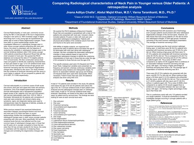
Comparing Radiological characteristics of Neck Pain in Younger versus Older Patients: A retrospective analysis
Jnana Aditya Challa, Abdul Majid Khan, and Varna Taranikanti
Publication Date: 5-2-2022
INTRODUCTION
Neck pain commonly occurs during the fifth or sixth decade of life due to degenerative changes in the spine. With increased usage of digital technology from a very young age we hypothesized an earlier age of onset of these degenerative changes. There have been no recent epidemiologic studies that investigated difference in radiological changes seen in older versus younger patients presenting with neck pain. Hence, this study is undertaken to analyze the variability in radiological changes seen in the cervical vertebrae between older (>50) versus younger (≤50) patients presenting with the chief complaint of neck pain. -

Pre and Postnatal Magnetic Resonance Imaging of Ventriculomegaly
Ryan Kelsch, Megan Moore, and Anant Krishnan
Publication Date: 4-2022
Purpose or Case Report: The purpose of this research was to analyze our institution’s large database of fetal magnetic resonance (MR) for cases of ventriculomegaly in order to understand trends in pre and postnatal MR.
Methods & Materials: In this retrospective study, 316 individual fetal MR exams from the past 10 years at our institution were reviewed. Of those, 86 patients had fetal MRs with findings of either ventriculomegaly or an ordering indication of ventriculomegaly. Our inclusion criteria of a diagnosis of ventriculomegaly (lateral ventricle measured at the trigone on coronal imaging of over 10mm) on fetal MR with a corresponding postnatal MR for that patient yielded 21 patients. Information extracted included degree of ventriculomegaly, and cause as determined by imaging. Correlation was performed via chart review to understand each patient's clinical outcome. Poor outcome was defined as permanent neurological deficits including seizures and developmental delay. The majority of the clinical outcome information was collected from the first few years of life.
Results: Of the 21 patients with ventriculomegaly with pre and postnatal MR imaging, the cause for ventriculomegaly was determined by prenatal imaging in 10 patients, by postnatal imaging in 4, while in 7 patients, a definite cause was not determined by the combination of prenatal and post-natal imaging. On prenatal imaging 7 fetuses had mild ventriculomegaly (10-12mm), 6 fetuses had moderate ventriculomegaly (>12-15mm) and 6 fetuses had severe ventriculomegaly (>15mm). Of the patients with mild ventriculomegaly, 5/7 had a normal neurological outcome while 2/7 have thus far had a poor neurological outcome. Of the patients with moderate ventriculomegaly, all 6, and similarly of the severe ventriculomegaly, 4/5 have thus far had a poor neurological outcome, with one patient not having enough clinical information thus far to determine outcome.
Conclusions: Our study demonstrates the utility of fetal MR in characterizing ventriculomegaly, with 48% (10/21) of patients receiving an etiology for the ventriculomegaly based on fetal MR findings. Our study also confirms previously reported studies that fetuses with mild ventriculomegaly more often have a normal neurological outcome (71%, 5/7) when compared to those with moderate to severe ventriculomegaly. -

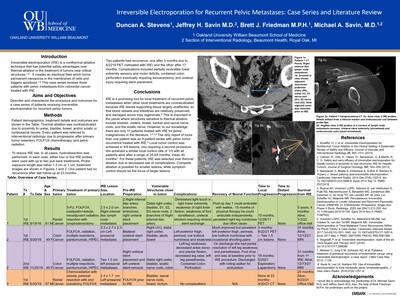
Irreversible Electroporation for Recurrent Pelvic Metastases: Case Series and Literature Review
Duncan A. Stevens, Jeffrey H. Savin, Brett J. Friedman, and Michael A. Savin
Publication Date: 9-23-2022
Purpose: Irreversible electroporation (IRE) is a nonthermal ablative technique that has potential safety advantages over thermal ablation in the treatment of patients with tumors near critical structures. It creates an electrical field that forms permanent nanopores in the membranes of cells and triggers apoptosis. This case series reviews three patients with pelvic metastases from colorectal cancer treated with IRE.
Materials and Methods: Two patients had rectal cancer, and one had sigmoid colon cancer. The mean age was 55 years, and there were two men and one woman. Thermal ablation was contraindicated because of proximity to the ureter, bladder, bowel, or sciatic or lumbosacral nerves. Every patient was referred to interventional radiology because of progression after primary tumor resection, FOLFOX (folinic acid, fluorouracil, and oxaliplatin) chemotherapy, and pelvic radiation. All patients were treated with NanoKnife IRE (AngioDynamics).
Results: To reduce IRE risk, hydrodissection was performed. In each case, either four or five IRE probes were used with up to two pull-back treatments. The probe exposure length was either 1.5 cm or 1 cm. One patient had no recurrence after the last follow-up at 23 months. Two patients had recurrence, one after 6 months (retreated with IRE) and the other after 17 months. Complications included partially reversible lower extremity sensory and motor deficits, contained colon perforation, and ureteral injury requiring stent placement.
Conclusions: IRE is a promising tool for local treatment of recurrent pelvic metastases when other local treatments are contraindicated. IRE leaves supporting tissue largely unaffected so that blood vessels and the intestines are relatively preserved, and damaged axons may regenerate. This is important in the pelvis, where sensitive structures include the bladder, ureters, bowel, lumbar and sacral nerve roots, and sciatic nerve. For these patients, IRE was selected over thermal ablation because of a decreased risk of complications. Complete ablation is possible for smaller lesions, but symptom control should be the focus for patients with larger lesions.
-

Irreversible Electroporation for Recurrent Pelvic Metastases: Case Series and Literature Review
Duncan A. Stevens, Jeffrey H. Savin, Brett J. Friedman, and Michael A. Savin
Publication Date: 5-2-2022
INTRODUCTION
Irreversible electroporation (IRE) is a nonthermal ablative technique that has potential safety advantages over thermal ablation in the treatment of tumors near critical structures. It creates an electrical field which forms permanent nanopores in the membranes of cells and triggers apoptosis. This case series reviews three patients with pelvic metastases from colorectal cancer treated with IRE.
Printing is not supported at the primary Gallery Thumbnail page. Please first navigate to a specific Image before printing.