


-

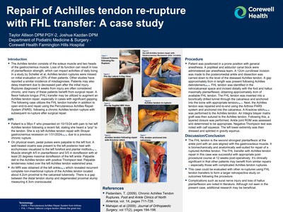
Repair of Achilles Tendon Re-Rupture with FHL Transfer: A Case Study
Taylor Allison and Joshua Kazdan
Publication Date: 3-13-2025
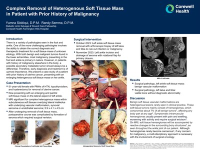
The Achilles tendon consists of the soleus muscle and two heads of the gastrocnemius muscle. Loss of its function can result in loss of plantarflexion strength, which can impact activities of daily living. In a study by Scheller et al, Achilles tendon ruptures were missed on initial evaluation on 25% of their patients. Other studies have reported a similar incidence of misdiagnoses. Patients may also delay treatment due to decreased pain after the initial injury. Ruptures diagnosed 4 weeks from injury are often considered chronic, and many of these patients benefit from surgical repair. A flexor hallucis longus (FHL) transfer may be utilized to augment an Achilles tendon repair, especially in cases with significant gapping. The following case utilizes the FHL tendon transfer in addition to open end-to-end repair using the Percutaneous Achilles Repair System (PARS) following a chronic Achilles tendon rupture with subsequent re-rupture after surgical repair.
-

Pyogenic Tenosynovitis of Tibialis Anterior Tendon after Cat Bite
Daniel Blascak, David Sosnoski, and Aaron Seidman
Publication Date: 3-13-2025
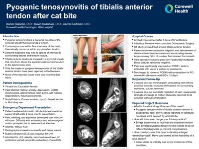
▪ Pyogenic tenosynovitis is a bacterial infection of the synovial sheath that surrounds a tendon.
▪ Commonly occurs within flexor tendons of the hand; theoretically can occur within any sheathed tendon.
▪ Delayed diagnosis may lead to spread of infection to surrounding tissues and tendon rupture.
▪ Tibialis anterior tendonis encased in asynovial sheath that runs from above the superior extensor retinaculum to the talonavicular joint.
▪ Only five cases of pyogenic tenosynovitis of the tibialis anterior tendon have been reported in the literature.
▪ None of the reported cases were due to animal bite injury.
-

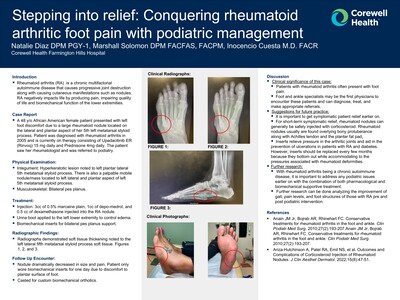
Stepping into Relief: Conquering Rheumatoid Arthritic Foot Pain with Podiatric Management
Natalie Diaz, Marshall Solomon, and Inocencio Cuesta
Publication Date: 3-13-2025
Rheumatoid arthritis (RA) is a chronic multifactorial autoimmune disease that causes progressive joint destruction along with causing cutaneous manifestations such as nodules. RA negatively impacts life by producing pain, impairing quality of life and biomechanical function of the lower extremities.
-

Kaposi Sarcoma: Case Study
Mohamed Elshebiny and Marshall G. Solomon
Publication Date: 3-13-2025
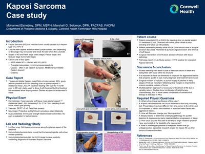
Kaposi Sarcoma (KS) is a vascular tumor usually caused by a herpestype virus HHV-8
Lesions often appear as flat or raised purple-colored, and depending on the stage may be nodular and break the skin when they ulcerate.
Stages of KS are Patch stage (early stage), Plaque stage, and Nodular stage (tumor/late stage).
Can be one of four types:
–AIDS related KS – infected with HIV (AKS)
– Transplant-associated – in immunosuppressed pts
Classic – often in pts Eastern-European, Mediterranean/Middle Eastern descent
– Endemic
-

Complications and Formation of Sinus Tracts Due To Presence of Lead From Foreign Bodies Insoft Tissue: A Case Report
Peter J. Godoy and Marshall G. Solomon
Publication Date: 3-13-2025
A cornerstone of lower extremity limb salvage is to avoid development of wound complicationsinvolved in soft tissue infections .
Predicting probability of sinus tract development due to presence of lead in soft tissues from foreign bodies. Inthis particular case a bulletfragment from a previous gunshot wound was noted to be the catalyst disrupting multiple biologic processes and impairing the function of soft tissues to heal. Predicting a level of contamination in this particular case leadbelieved to cause oxidative stress and inflammation and prompt excisional removal canavoid sinus tract formations and complications.
This study presents a unique case that demonstratesbeneficial patient care when a initial suspicion for wound contamination via lead and Swift intervention can lowerprobability of complications of wound healing.
-

The Diagnosis of Polyarteritis Nodosa in the Setting of Lower Extremity Lesions: A Case Report
Eric Li and Jihan Toma
Publication Date: 3-13-2025
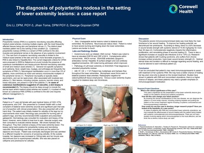
Polyarteritis nodosa (PAN) is a systemic necrotizing vasculitis affecting medium-sized arteries, involving multiple organs, with the most frequently affected tissues being skin and peripheral nerves (1). The distinct pearl neckless pattern led to the naming of this condition (2). Cutaneous polyarteritis nodosa (CPAN) is a variant of PAN confined to the skin, muscles and peripheral nerves in the absence of any systemic involvement (1). This rare form of systemic necrotizing vasculitis is limited to the medium-sized arteries of the skinand has a more favorable prognosis (1). PAN is also linked to Hepatitis B(2).The current diagnostic criteria for CPAN were proposed in 2009 by Nakamura et al.and include the presence of cutaneous manifestations and biopsy-proven fibrinoid necrotizing vasculitis of small and medium sized arteries (1). General non-specific symptoms such asthenia, fever, weight loss, myalgia, and arthralgia are frequently the symptoms of PAN (1). Neurological manifestation occur in more than 2/3 of patients, more commonlyas motor and sensory mononeuritis multiplex of the peripheral nerves (1). Peripheral neuropathy is typically distal, asymmetric, and can be rapid-onset, often associated with localized skin edema (1). Skin lesions, including nodules, purpura, necrotic ulcers, and livedo reticularis, are present in half of the patients (1). In cases of cutaneous manifestations suggestive of vasculitis, a skin biopsy is recommended (1). The biopsy should be deep enough to include the dermal layer where medium-sized arteries are located (1). A subset of PAN, known as cutaneous PAN (CPAN) is confined to the skin and requires different management (1).
-

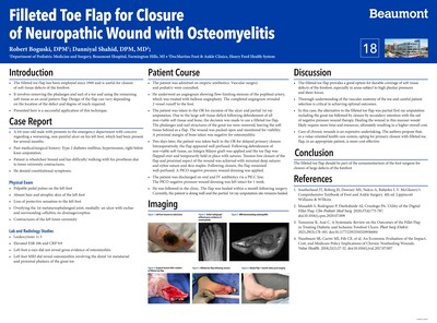
The Curious Case of Ainhum Dactolysis Spontanea
Jesse Miller, Michele Bertele-Semma, and Randy Semma
Publication Date: 3-13-2025
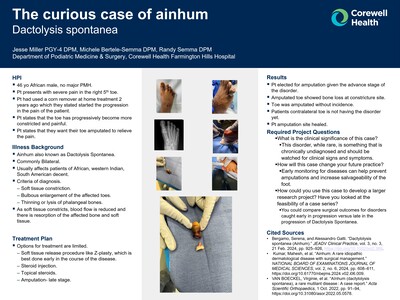
HPI
- 46 yoAfrican male, no major PMH.
- Pt presents with severe pain in the right 5thtoe.
- Pt had used a corn remover at home treatment 2 years ago which they stated started the progression in the pain of the patient.
- Pt states that the toe has progressively become more constricted and painful.
- Pt states that they want their toe amputated to relieve the pain.
-

Case Report of Surgical Management for Charcot Foot Medial Column Beaming with STJ Arthrodesis
Prachi Patel and Randy Semma
Publication Date: 3-13-2025
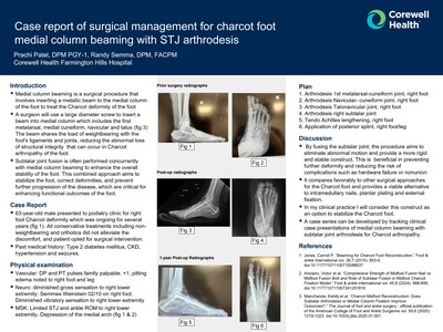
Medial column beaming is a surgical procedure that involves inserting a metallic beam to the medial column of the foot to treat the Charcot deformity of the foot.
A surgeon will use a large diameter screw to insert a beam into medial column which includes the first metatarsal, medial cuneiform, navicular and talus (fig 3) The beam shares the load of weightbearing with the foot's ligaments and joints, reducing the abnormal loss of structural integrity that can occur in Charcot arthropathy of the foot.
Subtalar joint fusion is often performed concurrently with medial column beaming to enhance the overall stability of the foot. This combined approach aims to stabilize the foot, correct deformities, and prevent further progression of the disease, which are critical for enhancing functional outcomes of the foot.
-

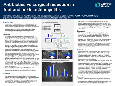
Antibiotics vs Surgical Resection in Foot and Ankle Osteomyelitis
Travis Rich and Marshall G. Solomon
Publication Date: 3-13-2025
Osteomyelitis, a serious bone infection, is particularly challenging in diabetic patients due to poor vascularization and impaired immune response.1 Timely and effective treatment is essential to prevent severe complications, including limb loss or even death.2 This study aims to evaluate the likelihood of surgical excision or amputation versus antibiotic treatment alone in diabetic patients with bone biopsyconfirmed acute osteomyelitis of the lower extremity.
-

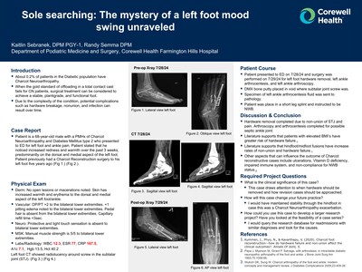
Sole searching: The mystery of a left foot mood swing unraveled Patient
Kaitlin Sebranek
Publication Date: 3-13-2025
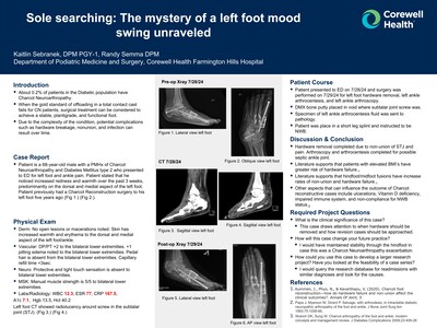
About 0.2% of patients in the Diabetic population have Charcot Neuroarthropathy.
When the gold standard of offloading in a total contact cast fails for CN patients, surgical treatment can be considered to achieve a stable, plantigrade, and functional foot.
Due to the complexity of the condition, potential complications such as hardware breakage, nonunion, and infection can result over time.
-

Repair of Achilles Tendon Re-Rupture with FHL Transfer: A Case Study
Taylor Allison and Joshua Kazdan
Publication Date: 5-9-2024
The Achilles tendon consists of the soleus muscle and two heads of the gastrocnemius muscle. Loss of its function can result in loss of plantarflexion strength, which can impact activities of daily living. In a study by Scheller et al, Achilles tendon ruptures were missed on initial evaluation on 25% of their patients. Other studies have reported a similar incidence of misdiagnoses. Patients may also delay treatment due to decreased pain after the initial injury. Ruptures diagnosed 4 weeks from injury are often considered chronic, and many of these patients benefit from surgical repair. A flexor hallucis longus (FHL) transfer may be utilized to augment an Achilles tendon repair, especially in cases with significant gapping. The following case utilizes the FHL tendon transfer in addition to open end-to-end repair using the Percutaneous Achilles Repair System (PARS) following a chronic Achilles tendon rupture with subsequent re-rupture after surgical repair.
-

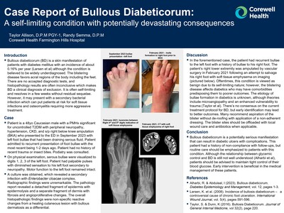
Case Report of Bullous Diabeticorum: A self-limiting condition with potentially devastating consequences
Taylor Allison and Randy Semma
Publication Date: 5-9-2024
Bullous diabeticorum (BD) is a skin manifestation of patients with diabetes mellitus with an incidence of about 0.16% per year (Larsen et al) although the condition is believed to be widely underdiagnosed. The blistering disease favors acral regions of the body including the feet. There are no accepted diagnostic tests, and histopathology results are often inconclusive which makes BD a clinical diagnosis of exclusion. It is often self-limiting and resolves in a few weeks without residual sequelae. However, it may present with a secondary bacterial infection which can put patients at risk for soft tissue infections and osteomyelitis requiring more aggressive treatment.
▪ Bullous diabeticorum (BD) is a skin manifestation of patients with diabetes mellitus with an incidence of about 0.16% per year (Larsen et al) although the condition is believed to be widely underdiagnosed. The blistering disease favors acral regions of the body including the feet. There are no accepted diagnostic tests, and histopathology results are often inconclusive which makes BD a clinical diagnosis of exclusion. It is often self-limiting and resolves in a few weeks without residual sequelae. However, it may present with a secondary bacterial infection which can put patients at risk for soft tissue infections and osteomyelitis requiring more aggressive treatment.
-

Septic First Metatarsophalangeal Joint Arthrodesis with Alternative Mini-Rail Fixation
Brenna Barker, Morgan Gascoyne, and Randy Semma
Publication Date: 5-9-2024
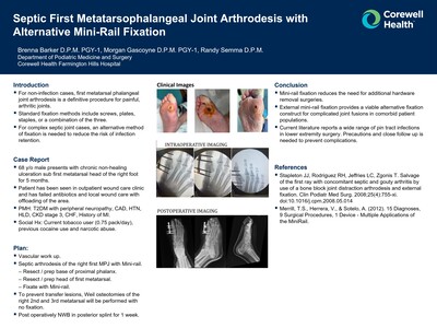
For non-infection cases, first metatarsal phalangeal joint arthrodesis is a definitive procedure for painful, arthritic joints.
Standard fixation methods include screws, plates, staples, or a combination of the three.
For complex septic joint cases, an alternative method of fixation is needed to reduce the risk of infection retention.
-

Stepping Into Relief: Conquering Rheumatoid Arthritic Foot Pain with Podiatric Management
Natalie Diaz, Marshall Solomon, and Inocencio Cuesta
Publication Date: 5-9-2024
Rheumatoid arthritis (RA) is a chronic multifactorial autoimmune disease that causes progressive joint destruction along with causing cutaneous manifestations such as nodules. RA negatively impacts life by producing pain, impairing quality of life and biomechanical function of the lower extremities.
-

Complications and Formation of Sinus Tracts Due to Presence of Lead From Foreign Bodies in Soft Tissue: A Case Report
Peter J. Godoy and Marshall G. Solomon
Publication Date: 5-9-2024
A cornerstone of lower extremity limb salvage is to avoid development of wound complications involved in soft tissue infections . Predicting probability of sinus tract development due to presence of lead in soft tissues from foreign bodies. In this particular case a bullet fragment from a previous gunshot wound was noted to be the catalyst disrupting multiple biologic processes and impairing the function of soft tissues to heal. Predicting a level of contamination in this particular case lead believed to cause oxidative stress and inflammation and prompt excisional removal can avoid sinus tract formations and complications. This study presents a unique case that demonstrates beneficial patient care when a initial suspicion for wound contamination via lead and Swift intervention can lower probability of complications of wound healing.
-

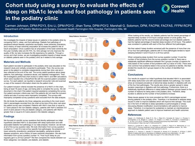
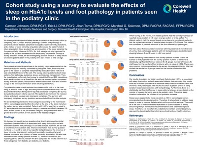
Cohort Study Using a Survey to Evaluate the Effects of Sleep on Hba1c Levels and Foot Pathology in Patients Seen in the Podiatry Clinic
Carmen Johnson, Eric Li, Jihan Toma, and Marshall G. Solomon
Publication Date: 5-9-2024
We investigate the impacts of sleep issues on patients in the podiatry clinic by comparing HbA1c, sleep, and foot pathology. Diabetic foot pathology of peripheral arterial disease, peripheral neuropathy, lower extremity ulcerations, and a history of lower extremity amputation all increase the patient's risk of future amputation. Once a patient has an amputation of the lower extremity the five-year mortality rates are 40-70%. So, limb salvage not only improves the quality of life, but also increases the life expectancy for patients. Through a survey and chart review, we are investigating possible impacts of sleep issues on diabetic patients seen in the podiatry clinic as it relates to limb salvage
-

The Diagnosis of Polyarteritis Nodosa in the Setting of Lower Extremity Lesions: A Case Report
Eric Li, Jihan Toma, and George Gopoian
Publication Date: 5-9-2024
Polyarteritis nodosa (PAN) is a systemic necrotizing vasculitis affecting medium-sized arteries, involving multiple organs, with the most frequently affected tissues being skin and peripheral nerves (1). The distinct pearl neckless pattern led to the naming of this condition (2). Cutaneous polyarteritis nodosa (CPAN) is a variant of PAN confined to the skin, muscles and peripheral nerves in the absence of any systemic involvement (1). This rare form of systemic necrotizing vasculitis is limited to the medium-sized arteries of the skin and has a more favorable prognosis (1). PAN is also linked to Hepatitis B(2). The current diagnostic criteria for CPAN were proposed in 2009 by Nakamura et al.and include the presence of cutaneous manifestations and biopsy-proven fibrinoid necrotizing vasculitis of small and medium sized arteries (1). General non-specific symptoms such asthenia, fever, weight loss, myalgia, and arthralgia are frequently the symptoms of PAN (1). Neurological manifestation occur in more than 2/3 of patients, more commonly as motor and sensory mononeuritis multiplex of the peripheral nerves (1). Peripheral neuropathy is typically distal, asymmetric, and can be rapid-onset, often associated with localized skin edema (1). Skin lesions, including nodules, purpura, necrotic ulcers, and livedo reticularis, are present in half of the patients (1). In cases of cutaneous manifestations suggestive of vasculitis, a skin biopsy is recommended (1). The biopsy should be deep enough to include the dermal layer where medium-sized arteries are located (1). A subset of PAN, known as cutaneous PAN (CPAN) is confined to the skin and requires different management (1).
-

The Curious Case of Ainhum Dactolysis Spontanea
Jesse Miller, Michele Bertele-Semma, and Randy Semma
Publication Date: 5-9-2024
46 yo African male, no major PMH.
Pt presents with severe pain in the right 5th toe.
Pt had used a corn remover at home treatment 2 years ago which they stated started the progression in the pain of the patient.
Pt states that the toe has progressively become more constricted and painful.
Pt states that they want their toe amputated to relieve the pain.
-

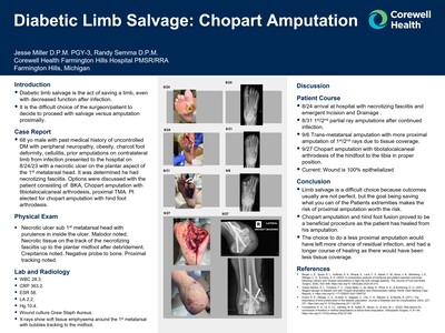
Diabetic Limb Salvage: Chopart Amputation
Jesse Miller and Randy Semma
Publication Date: 5-9-2024
Diabetic limb salvage is the act of saving a limb, even with decreased function after infection.
It is the difficult choice of the surgeon/patient to decide to proceed with salvage versus amputation proximally.
-

Case Report of Surgical Management for Charcot Foot Medial Column Beaming with STJ Arthrodesis
Prachi Patel and Randy Semma
Publication Date: 5-9-2024
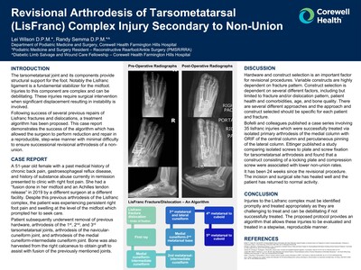
Medial column beaming is a surgical procedure that involves inserting a metallic beam to the medial column of the foot to treat the Charcot deformity of the foot.
A surgeon will use a large diameter screw to insert a beam into medial column which includes the first metatarsal, medial cuneiform, navicular and talus (fig 3) The beam shares the load of weightbearing with the foot's ligaments and joints, reducing the abnormal loss of structural integrity that can occur in Charcot arthropathy of the foot.
Subtalar joint fusion is often performed concurrently with medial column beaming to enhance the overall stability of the foot. This combined approach aims to stabilize the foot, correct deformities, and prevent further progression of the disease, which are critical for enhancing functional outcomes of the foot.
-

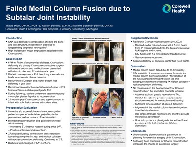
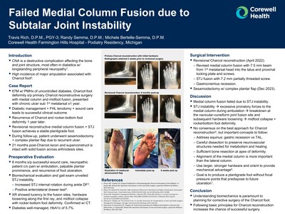
Failed Medial Column Fusion due to Subtalar Joint Instability
Travis Rich, Randy Semma, and Michele Bertelle-Semma
Publication Date: 5-9-2024
CNA is a destructive complication affecting the bone and joint structure, most often in diabetics w/ longstanding peripheral neuropathy.
High incidence of major amputation associated with Charcot foot.
-

Antibiotics Vs Surgical Resection in Foot and Ankle Osteomyelitis
Travis Rich and Marshall G. Solomon
Publication Date: 5-9-2024
Osteomyelitis, a serious bone infection, is particularly challenging in diabetic patients due to poor vascularization and impaired immune response.1 Timely and effective treatment is essential to prevent severe complications, including limb loss or even death.2 This study aims to evaluate the likelihood of surgical excision or amputation versus antibiotic treatment alone in diabetic patients with bone biopsyconfirmed acute osteomyelitis of the lower extremity.
-

Sole Searching: The Mystery of a Left Foot Mood Swing Unraveled
Kaitlin Sebranek and Randy Semma
Publication Date: 5-9-2024
About 0.2% of patients in the Diabetic population have Charcot Neuroarthropathy.
When the gold standard of offloading in a total contact cast fails for CN patients, surgical treatment can be considered to achieve a stable, plantigrade, and functional foot.
Due to the complexity of the condition, potential complications such as hardware breakage, nonunion, and infection can result over time.
Printing is not supported at the primary Gallery Thumbnail page. Please first navigate to a specific Image before printing.
